
Guides7-minute readFor medtechs reviewing Blood Bank and Hematology
Blood bank and hematology for the Prometric lab exam: 40% of the SCFHS technician paper
Blood bank and hematology review for the Prometric lab exam: how SCFHS and DHA list them, how to reason through ABO and morphology, and a two-week block.
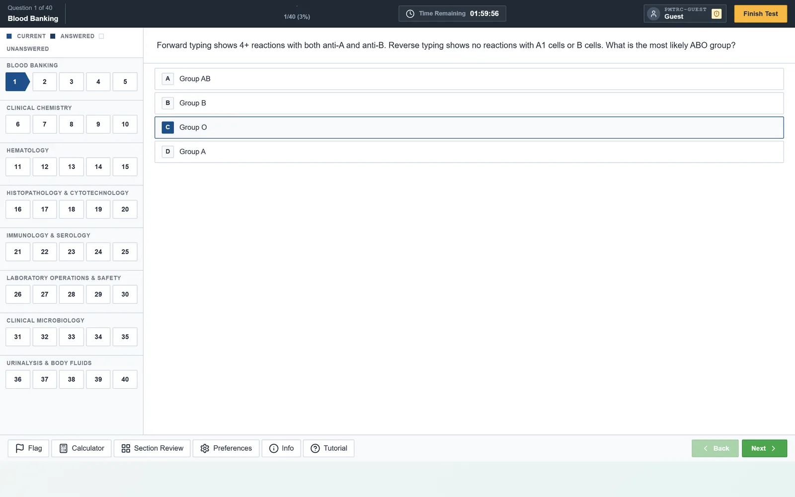
Blood bank and hematology are the two disciplines most medtechs think they already know, because they run them at the bench. The exam asks for the reasoning behind the bench: why a reverse type fails, which cell rules out an antibody, what a smear tells you before the analyser does. This guide shows how to review blood bank and hematology as patterns, with worked examples you can check yourself.
We keep the authorities apart, as the rest of this blog does. The weights below are published figures that can change, and where an authority publishes no weights we say so.
How much of the Prometric exam is blood bank and hematology?
The answer depends on the authority, and only SCFHS and the SLLE publish weights. The SCFHS Laboratory Technician blueprint gives Blood Bank 20% and Hematology 20% of a 150-item, 3-hour paper, which is about 60 items together. SCFHS adds that weights may differ by up to 5% in each category and are subject to change at any time.
The Laboratory Specialist exam (the SLLE) lists Blood Bank 15-20% and Hematology 15-20% among its sections. DHA's coverage lists for both its technician and technologist papers name blood bank and hematology, but DHA publishes no weights, so we will not invent any.
Whichever list is yours, treat the pair as a priority. Our SCFHS exam guide lays out all three SCFHS papers, and the time management guide shows how much clock these 60 items deserve. Our own bank holds 500 items in each, and the product's Full Mock draws 18 items from each of Blood Banking and Hematology. That blend is the product's design, not any authority's paper.
Why is blood bank a logic problem?
Blood bank rewards a fixed order of thinking, so learn the order and the facts attach themselves. Start with what the patient's cells carry (forward group), then what the patient's plasma carries (reverse group), and check that the two agree.
Discrepancies are a favourite exam topic. Work through them in order: check for a clerical error, repeat the test, then consider weak or missing antibodies (newborns, the elderly, immune deficiency), a cold autoantibody, rouleaux, or an A subgroup. The exam rarely wants a memorised list. It wants the next sensible step.
Rh follows the same discipline: D-positive or D-negative is a statement about one antigen, and a weak D result needs its own follow-up rather than a guess.
How do antibodies, compatibility testing and reactions fit together?
Antibody work is elimination, and compatibility testing is the safety net after it. Run the screen, then the panel. Cross out (rule out) an antibody when a panel cell that carries the antigen on a homozygous gene shows no reaction, because dosage can hide a heterozygous cell.
Next, use the rule of three: to call an antibody, you want at least three antigen-positive cells reacting and three antigen-negative cells not reacting. Check the autocontrol and the direct antiglobulin test (DAT) too. Think temperature as well: IgM antibodies such as anti-I and anti-M react in the cold, while clinically significant IgG antibodies in Rh, Kell, Duffy and Kidd react at the antiglobulin phase.
For a suspected transfusion reaction, the order is stop the transfusion, keep the line open, check patient and unit identification, inspect a post-reaction sample for haemolysis and run a DAT. Know the pattern for each reaction: acute haemolytic (usually an ABO error), febrile non-haemolytic, allergic or anaphylactic, TRALI and TACO.
Hemolytic disease of the fetus and newborn (HDFN) follows from maternal IgG crossing the placenta. A D-negative mother with a D-positive baby is the classic setup for Rh immune globulin.
| Topic | Core idea | How to drill |
|---|---|---|
| ABO and Rh typing | Forward and reverse must agree | Write out both groups for 10 cases, then add one discrepancy to each |
| Antibody panel | Rule out on homozygous cells, then rule of three | Redo the same panel with a different antigram until it takes under 5 minutes |
| Compatibility testing | Match ABO first, add antiglobulin when antibodies are present | Explain each phase aloud in one sentence |
| Transfusion reactions | Stop, identify, inspect, DAT | Make a one-line card per reaction type |
| HDFN | Maternal IgG against a fetal antigen | Trace D-negative mother, D-positive baby, from first pregnancy onward |
| Minor blood groups | Know which are IgG and clinically significant | Learn the five or six that cause reactions, not every system |
How should you study hematology: morphology and disorders?
Start every anaemia question from the MCV, then look at the smear, then pick the confirming test. That order cuts the options to two or three in seconds.
Microcytic anaemias are iron deficiency, thalassemia, anaemia of chronic disease and sideroblastic anaemia. Macrocytic anaemias split into megaloblastic (B12 or folate lack, hypersegmented neutrophils) and non-megaloblastic. Pair leukaemias with their signature finding: the Philadelphia chromosome in CML, Auer rods and t(15;17) in acute promyelocytic leukaemia, smudge cells in CLL.
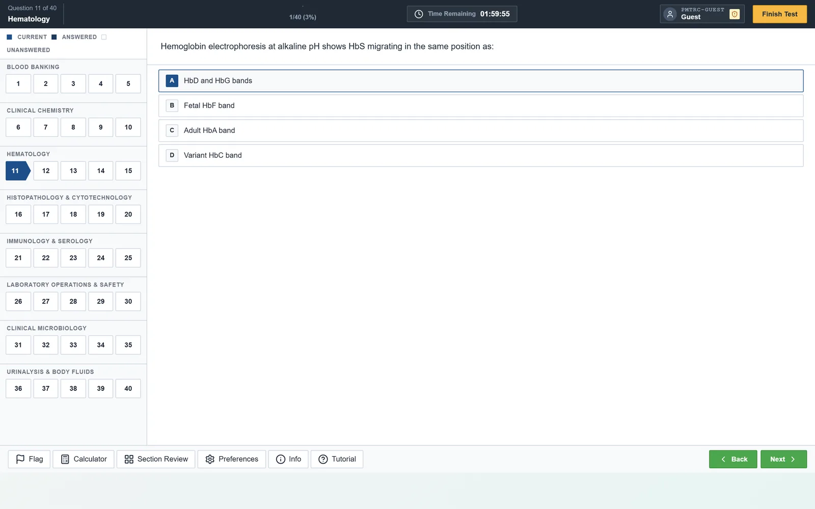
For that item, the fact to hold is that at alkaline pH, HbS migrates with HbD and HbG, and HbC migrates with HbE, HbA2 and HbO. Acid citrate agar is how you tell those groups apart.
What do you need to know about hemostasis?
Hemostasis is a map. The PT watches the extrinsic and common pathways (factor VII is the key extrinsic factor), and the aPTT watches the intrinsic and common pathways. Learn which test each drug moves: warfarin lengthens the PT (reported as INR) and unfractionated heparin lengthens the aPTT.
Read the pair of results as a pattern. A prolonged aPTT with a normal PT points toward the intrinsic pathway, such as haemophilia A (factor VIII) or B (factor IX) or von Willebrand disease. A prolonged PT with a normal aPTT points toward factor VII lack or early warfarin effect. Both prolonged suggests a common-pathway problem, liver disease or DIC.
A mixing study separates a factor deficiency (the prolongation corrects) from an inhibitor (it does not). In DIC, expect low platelets, low fibrinogen, raised D-dimer and prolonged times. Platelet disorders get their own logic: count first, then function.
What does a two-week blood bank and hematology block look like?
Spend the first week on blood bank and the second on hematology and hemostasis, with a short drill every day. The plan below assumes about two hours a day and is meant to be scaled to your shifts.
- Days 1-3: ABO, Rh and discrepancies. Do one mixed set of 100 Blood Banking items and write the reason for every miss.
- Days 4-5: antibody panels, compatibility testing, then transfusion reactions and HDFN.
- Day 6: the flashcards for blood banking, then a timed random pool.
- Day 7: rest, or review only the cards you missed.
- Days 8-10: anaemia by MCV, then white-cell disorders and leukaemias.
- Days 11-12: hemostasis, using the PT and aPTT map and the common drugs.
- Days 13-14: a timed Hematology set, then a mixed set across both disciplines.
Each discipline has four fixed sets of 100 items plus a random pool drawn from all 500. Use a fixed set for a baseline, then the random pool to see whether the weak spots moved. Fit this block into the wider eight-week study plan, and keep the clinical chemistry block next in line. A free demo shows the exam room before you commit any time.
Frequently asked questions
How much of the Prometric exam is blood bank?
It depends on the authority. The SCFHS Laboratory Technician blueprint gives blood bank 20%, with a possible shift of up to 5%. The SLLE lists 15-20%. DHA names blood bank on its coverage lists but publishes no weight. Check your own authority's current document, because these figures can change.
What is the fastest way to learn cell morphology?
Learn it in families, not one cell at a time. Group red cell shapes by what causes them (target cells, spherocytes, schistocytes, sickle cells) and pair each with the disease it signals. Then review images against a short written description. Flashcards with a picture on one side suit this well.
Do I need every minor blood group system?
No. Learn the systems that cause clinically significant antibodies, mainly Rh, Kell, Duffy, Kidd and MNS, plus the common cold-reacting ones such as Lewis, I and P1. Know each antibody's typical temperature and whether it causes transfusion reactions or HDFN. Rare systems matter far less than the reasoning steps.
Is blood bank harder than hematology?
Neither has published pass-rate data, so no source can answer that. Most reviewees find blood bank more procedural and hematology more visual. Practise each in its own style: step-by-step working for blood bank, pattern recognition under a clock for hematology.
Sources
From the publisher
Practise the lab disciplines first.
TerumoEdge Gulf MLS is a practice simulator for the Gulf laboratory licensing exams: timed sessions in eight lab disciplines, an adjustable clock, and a full explanation for every item instead of a bare answer key.
- 4,000 items, 500 in each of eight lab disciplines
- A 100-item blended mock, reshuffled each attempt
- A full English explanation for every item, with the reason each wrong option is wrong
- A lab science reviewer and flashcards beside the question bank
Try the free Gulf MLS simulator demo Forty items across all eight lab disciplines. No account needed.
About this article
Written by the TerumoEdge editorial team, who build the TerumoEdge Gulf MLS Ex-Sim. Exam facts are checked against the published documents of SCFHS, the Dubai Health Authority, Qatar's Department of Healthcare Professions and NHRA Bahrain, and every article lists its sources. How we write and check our articles.
First published .